The effect of surface microtopography of poly(dimethylsiloxane) on protein adsorption, platelet and cell adhesion
Colloids and Surfaces B: Biointerfaces,
2009,
71,
275-281.
文章链接:http://dx.doi.org/10.1016/j.colsurfb.2009.02.018
The effect of surface microtopography of poly(dimethylsiloxane) on protein adsorption, platelet and
发布日期:2009-01-25
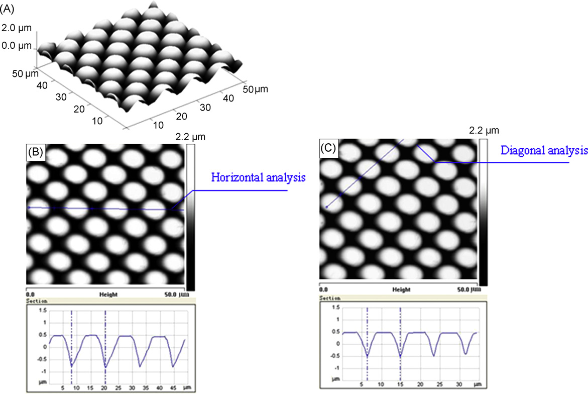
Chemical homogeneous poly(dimethylsiloxane) (PDMS) surface with dot-like protrusion pattern was used to investigate the individual effect of surface microtopography on protein adsorption and subsequent biological responses. Fibrinogen (Fg) and fibronectin (Fn)were chosen as model proteins due to their effect on platelet and cell adhesion, respectively. Fg labeled with 125I and fluorescein isothiocyanate (FITC) was
used to study its adsorption on flat and patterned surfaces. Patterned surface has a 46% increase in the adsorption of Fg when compared with flat surface. However, the surface area of the patterned surfacewas only 8%larger than that of the flat surface. Therefore, the increase in the surface areawas not the only factor responsible for the increase in protein adsorption. Clear fluorescent pattern was visualized on patterned surface, indicating that adsorbed Fg regularly distributed and adsorbed most on the flanks and valleys of the protrusions. Such distribution and local enrichment of Fg presumably caused the specific location of platelets adhered from platelet-rich plasma (PRP) and flowing whole blood (FWB) on patterned surface. Furthermore, the different combination of surface topography and pre-adsorbed Fn could influence the adhesion of L929 cells. The flat surface with pre-adsorbed Fnwas the optimum substrate while the virgin patterned surface was the poor substrate in terms of L929 cells spread.